Advertisement: The advertising company is responsible for the content of this page.


LipiScan
Dynamic meibomian gland imaging
The first meibomian gland imager developed for efficiency and flexibility in clinical use.
The LipiSCAN imaging system
With a small footprint and user-friendly design, the LipiScan ™ for Dynamic Meibomian Gland Imaging ™ (DMI) was developed to make high-resolution meibography available for every practice.
The stable and at the same time light device was developed for the optimization of the work processes and the easy integration into the busy practice. Both eyelids can be displayed in just about a minute.
How the LipiSCAN system works
Maximum acceleration of the workflow with fast imaging of the meibomian glands
Low space requirement and low weight for optimal versatility
Fast and intuitive operation for seamless integration into the usual clinical workflow
Favorable price to equip even small practices and various workplaces with the device
Creates a high-resolution image of the meibomian gland structure
Images can be exported as PDF
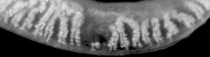
DYNAMIC MEIBOM IMAGING
High resolution image of the meibomian glands
Dynamic lighting
The surface lighting is realized by different light sources, which minimize disturbing reflections.
Adaptive fluoroscopy
Different light intensity over the surface of the lighting unit compensates for the differences in the thickness of the eyelid in different patients.
Dual mode DMI
A combination of dynamic lighting and adaptive fluoroscopy allows a significantly improved representation of the meibomian gland structure
